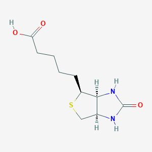
General Information of MET (ID: META00129)
Name Biotin
Synonyms   Click to Show/Hide Synonyms of This Metabolite
(+)-Biotin; (+)-cis-Hexahydro-2-oxo-1H-thieno[3,4]imidazole-4-valerate; (+)-cis-Hexahydro-2-oxo-1H-thieno[3,4]imidazole-4-valeric acid; (3AS,4S,6ar)-hexahydro-2-oxo-1H-thieno[3,4-D]imidazole-4-valerate; (3AS,4S,6ar)-hexahydro-2-oxo-1H-thieno[3,4-D]imidazole-4-valeric acid; -(+)-Biotin; 1SWK; 1SWN; 1SWR; 5-(2-Oxohexahydro-1H-thieno[3,4-D]imidazol-4-yl)pentanoate; 5-(2-Oxohexahydro-1H-thieno[3,4-D]imidazol-4-yl)pentanoic acid; Biocur brand OF biotin; Biodermatin; Bioepiderm; Biokur; Bios H; Bios II; Biotin biocur brand; Biotin dermapharm brand; Biotin gelfert; Biotin hermes; Biotin hermes brand; Biotin medopharm brand; Biotin ratiopharm; Biotin ratiopharm brand; Biotin roche brand; Biotin simons brand; Biotin strathmann brand; Biotin ziethen brand; Biotin-ratiopharm; Biotina; Biotine; Biotine roche; Biotinratiopharm; Biotinum; Coenzyme R; D(+)-Biotin; D-(+)-Biotin; D-Biotin; D-Biotin factor S; Deacura; Dermapharm brand OF biotin; E+b pharma brand OF biotin; Factor S; Factor S (vitamin); Gabunat; Gelfert, biotin; H, Vitamin; Hermes brand OF biotin; Hermes, biotin; Hexahydro-2-oxo-1H-thieno(3,4-D)imidazole-4-pentanoate; Hexahydro-2-oxo-1H-thieno(3,4-D)imidazole-4-pentanoic acid; Hexahydro-2-oxo-[3as-(3aa,4b,6aa)]-1H-thieno[3,4-D]imidazole-4-pentanoate; Hexahydro-2-oxo-[3as-(3aa,4b,6aa)]-1H-thieno[3,4-D]imidazole-4-pentanoic acid; Hexahydro-2-oxo-[3as-(3alpha,4beta,6alpha)]-1H-thieno[3,4-D]imidazole-4-pentanoate; Hexahydro-2-oxo-[3as-(3alpha,4beta,6alpha)]-1H-thieno[3,4-D]imidazole-4-pentanoic acid; Lutavit H2; Medea brand OF biotin sodium salt; Medebiotin; Medobiotin; Medopharm brand OF biotin; Meribin; Ratiopharm brand OF biotin; Roche brand OF biotin; Roche, biotine; Rombellin; Rovimix H 2; Simons brand OF biotin; Strathmann brand OF biotin; Vitamin H; Vitamin b7; Vitamin-H; Ziethen brand OF biotin; cis-(+)-Tetrahydro-2-oxothieno[3,4]imidazoline-4-valerate; cis-(+)-Tetrahydro-2-oxothieno[3,4]imidazoline-4-valeric acid; cis-Hexahydro-2-oxo-1H-thieno(3,4)imidazole-4-valerate; cis-Hexahydro-2-oxo-1H-thieno(3,4)imidazole-4-valeric acid; cis-Tetrahydro-2-oxothieno(3,4-D)imidazoline-4-valerate; cis-Tetrahydro-2-oxothieno(3,4-D)imidazoline-4-valeric acid; delta-(+)-Biotin; delta-Biotin; delta-Biotin factor S
Source Food;Escherichia Coli Metabolite;Yeast Metabolite;Food;Drug;Toxins/Pollutant;Cosmetic;Food additives;TCM Ingredients;Microbial
Structure Type   Biotin and derivatives  (Click to Show/Hide the Complete Structure Type Hierarchy)
Organoheterocyclic compounds
Biotin and derivatives
PubChem CID
171548
HMDB ID
HMDB0000030
Formula
C10H16N2O3S
Structure
<iframe style="width: 300px; height: 300px;" frameborder="0" src="https://embed.molview.org/v1/?mode=balls&cid=171548"></iframe>
3D MOL 2D MOL
  Click to Show/Hide the Molecular/Functional Data (External Links/Property/Function) of This Metabolite
KEGG ID
C00120
DrugBank ID
DB00121
ChEBI ID
15956
FooDB ID
FDB014510
ChemSpider ID
149962
METLIN ID
243
Physicochemical Properties Molecular Weight 244.31 Topological Polar Surface Area 104
XlogP 0.3 Complexity 298
Heavy Atom Count 16 Rotatable Bond Count 5
Hydrogen Bond Donor Count 3 Hydrogen Bond Acceptor Count 4
Function
Biotin is an enzyme co-factor present in minute amounts in every living cell. Biotin is also known as coenzyme R and vitamin H or B7. It occurs mainly bound to proteins or polypeptides and is abundant in liver, kidney, pancreas, yeast, and milk. Biotin has been recognized as an essential nutrient. Humans fulfill their biotin requirement through their diet through endogenous reutilization of biotin and perhaps through the capture of biotin generated in the intestinal flora. The utilization of biotin for covalent attachment to carboxylases and its reutilization through the release of carboxylase biotin after proteolytic degradation constitutes the 'biotin cycle'. Biotin deficiency is associated with neurological manifestations, skin rash, hair loss, and metabolic disturbances that are thought to relate to the various carboxylase deficiencies (metabolic ketoacidosis with lactic acidosis). It has also been suggested that biotin deficiency is associated with protein malnutrition, and that marginal biotin deficiency in pregnant women may be teratogenic. Biotin acts as a carboxyl carrier in carboxylation reactions. There are four biotin-dependent carboxylases in mammals: those of propionyl-CoA (PCC), 3-methylcrotonyl-CoA (MCC), pyruvate (PC), and acetyl-CoA carboxylases (isoforms ACC-1 and ACC-2). All but ACC-2 are mitochondrial enzymes. The biotin moiety is covalently bound to the epsilon amino group of a lysine residue in each of these carboxylases in a domain 60-80 amino acids long. The domain is structurally similar among carboxylases from bacteria to mammals. Evidence is emerging that biotin participates in processes other than classical carboxylation reactions. Specifically, novel roles for biotin in cell signalling, gene expression, and chromatin structure have been identified in recent years. Human cells accumulate biotin by using both the sodium-dependent multivitamin transporter and monocarboxylate transporter 1. These transporters and other biotin-binding proteins partition biotin to compartments involved in biotin signalling: cytoplasm, mitochondria, and nuclei. The activity of cell signals such as biotinyl-AMP, Sp1 and Sp3, nuclear factor (NF)-kappaB, and receptor tyrosine kinases depends on biotin supply. Consistent with a role for biotin and its catabolites in modulating these cell signals, greater than 2000 biotin-dependent genes have been identified in various human tissues. Many biotin-dependent gene products play roles in signal transduction and localize to the cell nucleus, consistent with a role for biotin in cell signalling. Posttranscriptional events related to ribosomal activity and protein folding may further contribute to the effects of biotin on gene expression. Finally, research has shown that biotinidase and holocarboxylase synthetase mediate covalent binding of biotin to histones (DNA-binding proteins), affecting chromatin structure; at least seven biotinylation sites have been identified in human histones. Biotinylation of histones appears to play a role in cell proliferation, gene silencing, and the cellular response to DNA repair. Roles for biotin in cell signalling and chromatin structure are consistent with the notion that biotin has a unique significance in cell biology.
Regulatory Network
Full List of Protein(s) Regulating This Metabolite
      GPCR secretin (GPCR-2)
            Glucagon receptor (GCGR) Click to Show/Hide the Full List of Regulating Pair(s):   1 Pair(s)
               Detailed Information Protein   Info click to show the details of this protein
               Regulating Pair Experim Info click to show the details of experiment for validating this pair [1]
                      Introduced Variation Knockout of Gcgr
                      Induced Change Biotin concentration: increase (FC = 1.6)
                      Summary Introduced Variation         Induced Change 
                      Disease Status Type 2 diabetes mellitus [ICD-11: 5A11]
                      Details It is reported that knockout of GCGR leads to the increase of biotin levels compared with control group.
      Hydrolases (EC 3)
            Leukotriene-C4 hydrolase (GGT1) Click to Show/Hide the Full List of Regulating Pair(s):   1 Pair(s)
               Detailed Information Protein   Info click to show the details of this protein
               Regulating Pair Experim Info click to show the details of experiment for validating this pair [2]
                      Introduced Variation Knockdown (siRNA) of GGT1
                      Induced Change Biotin concentration: increase
                      Summary Introduced Variation         Induced Change 
                      Disease Status Renal cell carcinoma [ICD-11: 2C90]
                      Details It is reported that knockdown of GGT1 leads to the increase of biotin levels compared with control group.
            Sulfatase sulf-1 (SULF1) Click to Show/Hide the Full List of Regulating Pair(s):   1 Pair(s)
               Detailed Information Protein   Info click to show the details of this protein
               Regulating Pair Experim Info click to show the details of experiment for validating this pair [3]
                      Introduced Variation Knockdown (shRNA) of SULF1
                      Induced Change Biotin concentration: decrease (FC = 0.30 / 0.36)
                      Summary Introduced Variation         Induced Change 
                      Disease Status Ovarian cancer [ICD-11: 2C73]
                      Details It is reported that knockdown of SULF1 leads to the decrease of biotin levels compared with control group.
      Transcription factor (TF)
            R2R3-MYB (AN2) Click to Show/Hide the Full List of Regulating Pair(s):   1 Pair(s)
               Detailed Information Protein   Info click to show the details of this protein
               Regulating Pair Experim Info click to show the details of experiment for validating this pair [4]
                      Introduced Variation Overexpression of AN2
                      Induced Change Biotin concentration: decrease (FC = 0.38)
                      Summary Introduced Variation         Induced Change 
                      Disease Status Healthy individual
                      Details It is reported that overexpression of AN2 leads to the decrease of biotin levels compared with control group.
References
1 Polyomic profiling reveals significant hepatic metabolic alterations in glucagon-receptor (GCGR) knockout mice: implications on anti-glucagon therapies for diabetes. BMC Genomics. 2011 Jun 1;12:281.
2 Impairment of gamma-glutamyl transferase 1 activity in the metabolic pathogenesis of chromophobe renal cell carcinoma. Proc Natl Acad Sci U S A. 2018 Jul 3;115(27):E6274-E6282.
3 Erratum to: Loss of HSulf-1 promotes altered lipid metabolism in ovarian cancer. Cancer Metab. 2014 Nov 4;2:24.
4 Comprehensive Influences of Overexpression of a MYB Transcriptor Regulating Anthocyanin Biosynthesis on Transcriptome and Metabolome of Tobacco Leaves. Int J Mol Sci. 2019 Oct 16;20(20):5123.

If you find any error in data or bug in web service, please kindly report it to Dr. Zhang and Dr. Mou.