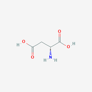
General Information of MET (ID: META00568)
Name D-Aspartic acid
Synonyms   Click to Show/Hide Synonyms of This Metabolite
(-)-Aspartic acid; (2R)-2-Aminobutanedioate; (2R)-2-Aminobutanedioic acid; (R)-2-Aminobutanedioate; (R)-2-Aminobutanedioic acid; (R)-2-Aminosuccinate; (R)-2-Aminosuccinic acid; (R)-Aspartic acid; 1-Amino-1,2-carboxyethane; Aspartate D-form; Aspartic acid; Aspartic acid D-form; D Aspartate; D Aspartic acid; D-(-)-Aspartic acid; D-Asparaginsaeure; D-Aspartate; DAS; Lopac-alpha-9256; Tocris-0213; delta-(-)-Aspartic acid; delta-Asparaginsaeure; delta-Aspartate; delta-Aspartic acid
Source Endogenous;Food;Drug
Structure Type   Amino acids, peptides, and analogues  (Click to Show/Hide the Complete Structure Type Hierarchy)
Organic acids and derivatives
Carboxylic acids and derivatives
Amino acids, peptides, and analogues
PubChem CID
83887
HMDB ID
HMDB0006483
Formula
C4H7NO4
Structure
<iframe style="width: 300px; height: 300px;" frameborder="0" src="https://embed.molview.org/v1/?mode=balls&cid=83887"></iframe>
3D MOL 2D MOL
  Click to Show/Hide the Molecular/Functional Data (External Links/Property/Function) of This Metabolite
KEGG ID
C00402
DrugBank ID
DB02655
ChEBI ID
17364
FooDB ID
FDB023933
ChemSpider ID
75697
METLIN ID
15
Physicochemical Properties Molecular Weight 133.1 Topological Polar Surface Area 101
XlogP -2.8 Complexity 133
Heavy Atom Count 9 Rotatable Bond Count 3
Hydrogen Bond Donor Count 3 Hydrogen Bond Acceptor Count 5
Function
D-Aspartic acid is the D-isomer of aspartic acid. Since its discovery in invertebrates, free D-aspartate (D-Asp) has been identified in a variety of organisms, including microorganisms, plants, and lower animals, mammals and humans. D-Asp in mammalian tissues is present in specific cells, indicating the existence of specific molecular components that regulate D-Asp levels and localization in tissues. In the rat adrenal medulla, D-Asp is closely associated with adrenaline-cells (A-cells), which account for approximately 80% of the total number of chromaffin cells in the tissue, and which make and store adrenaline. D-Asp appears to be absent from noradrenaline-cells (NA-cells), which comprise approximately 20% of the total number of chromaffin cells in the adrenal medulla, and which make and store noradrenaline. D-aspartate oxidase (EC 1.4.3.1, D-AspO), which catalyzes oxidative deamination of D-Asp, appears to be present only in NA-cells, suggesting that the lack of D-Asp in these cells is due to D-Asp oxidase-mediated metabolism of D-Asp. In the rat adrenal cortex, the distribution of D-Asp changes during development. It has been suggested that developmental changes in the localization of D-Asp reflects the participation of D-Asp in the development and maturation of steroidogenesis in rat adrenal cortical cells. D-Asp is involved in steroid hormone synthesis and secretion in mammals as well. D-Asp is synthesized intracellularly, most likely by Asp racemase (EC 5.1.1.13). Endogenous D-Asp apparently has two different intracellular localization patterns: cytoplasmic and vesicular. D-Asp release can occur through three distinct pathways: 1) spontaneous, continuous release of cytoplasmic D-Asp, which is not associated with a specific stimulus; 2) release of cytoplasmic D-Asp via a volume-sensitive organic anion channel that connects the cytoplasm and extracellular space; 3) exocytotic discharge of vesicular D-Asp. D-Asp can be released via a mechanism that involves the L-Glu transporter. D-Asp is thus apparently in dynamic flux at the cellular level to carry out its physiological function(s) in mammals.
Regulatory Network
Full List of Protein(s) Regulating This Metabolite
      Amino acid/auxin permease (AAAP)
            Sodium-coupled neutral amino transporter 10 (SLC38A10) Click to Show/Hide the Full List of Regulating Pair(s):   1 Pair(s)
               Detailed Information Protein   Info click to show the details of this protein
               Regulating Pair Experim Info click to show the details of experiment for validating this pair [1]
                      Introduced Variation Overexpression of SLC38A10
                      Induced Change D-Aspartic acid concentration: decrease
                      Summary Introduced Variation         Induced Change 
                      Disease Status Healthy individual
                      Details It is reported that overexpression of SLC38A10 leads to the decrease of D-aspartic acid levels compared with control group.
      Dicarboxylate/amino acid:cation symporter (DAACS)
            Excitatory amino acid transporter 3 (SLC1A1) Click to Show/Hide the Full List of Regulating Pair(s):   1 Pair(s)
               Detailed Information Protein   Info click to show the details of this protein
               Regulating Pair Experim Info click to show the details of experiment for validating this pair [2]
                      Introduced Variation Overexpression of SLC1A1
                      Induced Change D-Aspartic acid concentration: decrease
                      Summary Introduced Variation         Induced Change 
                      Disease Status Motor neuron disease [ICD-11: 8B60]
                      Details It is reported that overexpression of SLC1A1 leads to the decrease of D-aspartic acid levels compared with control group.
      Lyases (EC 4)
            Cystathionine gamma-lyase (CTH) Click to Show/Hide the Full List of Regulating Pair(s):   1 Pair(s)
               Detailed Information Protein   Info click to show the details of this protein
               Regulating Pair Experim Info click to show the details of experiment for validating this pair [3]
                      Introduced Variation Knockout of Cth
                      Induced Change D-Aspartic acid concentration: increase (FC = 2.10)
                      Summary Introduced Variation         Induced Change 
                      Disease Status Organic acid disorderss [ICD-11: 5C50]
                      Details It is reported that knockout of Cth leads to the increase of D-aspartic acid levels compared with control group.
      Pore-forming PNC peptide (PNC)
            Cellular tumor antigen p53 (TP53) Click to Show/Hide the Full List of Regulating Pair(s):   1 Pair(s)
               Detailed Information Protein   Info click to show the details of this protein
               Regulating Pair Experim Info click to show the details of experiment for validating this pair [4]
                      Introduced Variation Knockout of TP53
                      Induced Change D-Aspartic acid concentration: increase (Log2 FC=1.19)
                      Summary Introduced Variation         Induced Change 
                      Disease Status Colon cancer [ICD-11: 2B90]
                      Details It is reported that knockout of TP53 leads to the increase of D-aspartic acid levels compared with control group.
      Transferases (EC 2)
            SNF-related serine/threonine-protein kinase (SNRK) Click to Show/Hide the Full List of Regulating Pair(s):   1 Pair(s)
               Detailed Information Protein   Info click to show the details of this protein
               Regulating Pair Experim Info click to show the details of experiment for validating this pair [5]
                      Introduced Variation Knockdown (shRNA) of SNRK
                      Induced Change D-Aspartic acid concentration: decrease
                      Summary Introduced Variation         Induced Change 
                      Disease Status Healthy individual
                      Details It is reported that knockdown of SNRK leads to the decrease of D-aspartic acid levels compared with control group.
References
1 The neuronal and astrocytic protein SLC38A10 transports glutamine, glutamate, and aspartate, suggesting a role in neurotransmission. FEBS Open Bio. 2017 Apr 26;7(6):730-746.
2 Primary structure and functional characterization of a high-affinity glutamate transporter. Nature. 1992 Dec 3;360(6403):467-71.
3 Hydrogen Sulfide Is a Regulator of Hemoglobin Oxygen-Carrying Capacity via Controlling 2,3-BPG Production in Erythrocytes. Oxid Med Cell Longev. 2021 Feb 13;2021:8877691.
4 Integrative omics analysis of p53-dependent regulation of metabolism. FEBS Lett. 2018 Feb;592(3):380-393.
5 Sucrose Nonfermenting-Related Kinase Enzyme-Mediated Rho-Associated Kinase Signaling is Responsible for Cardiac Function. Circ Cardiovasc Genet. 2016 Dec;9(6):474-486.

If you find any error in data or bug in web service, please kindly report it to Dr. Zhang and Dr. Mou.